Figure 1.
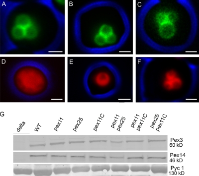
The H. polymorpha Pex11 family members. Fluorescence microscopy images of methanol-grown WT cells producing Pex11-GFP (A), Pex25-GFP (B), or Pex11C-GFP (C). All three proteins are localized to peroxisomes. (D–F) Fluorescence microscopy images of pex11 pex25 (D), pex11 pex11C (E), and pex25 pex11C cells (F) producing DsRed-SKL to mark the peroxisomal matrix. Cells were grown on glycerol/methanol mixtures. The DsRed-SKL fluorescence does not completely fill the matrix of the peroxisomes because of the presence of alcohol oxidase crystal inside the peroxisomes. Bar, 1 µm. Images were taken by wide-field fluorescence microscopy. The cell contour is indicated in blue. (G) Western blots showing the levels of Pex3 and Pex14 proteins in WT and various deletion strains. Cells were grown for 16 h on glycerol/methanol. Equal amounts of protein were loaded per lane. The first lane shows the negative controls of the corresponding deletion strain (i.e., pex3 and pex14). The pyruvate carboxylase (Pyc1) blot is added as loading control.

