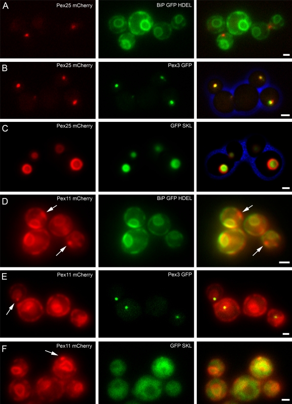
Figure 6.
Peroxisomes are formed in pex11 pex25 cells upon reintroduction of PEX25, but not upon reintroduction of PEX11. (A–C) pex11 pex25 PAOX PEX25-mCherry cells were shifted from glucose/ammonium sulfate to glycerol/methanol-containing media. Pex25-mCherry fluorescence is shown in the images in the left panels (in red). Cells were grown for 4 (A and B) or 16 h (C). (D–F) pex11 pex25 PAOX PEX11-mCherry cells were shifted from glucose/ammonium sulfate to glycerol/methanol medium. Cells were grown for 4 (D and E) or 16 h (F). The images at the right show the merged fluorescence images as well as the cell walls in blue. Bar, 1 µm.

