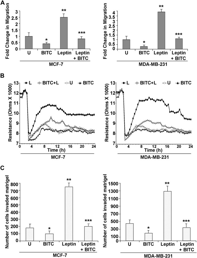
Fig. 2.
BITC inhibits leptin-induced migration and invasion of breast carcinoma cells. (A) Breast cancer cells (MCF-7 and MDA-MB-231) were subjected to scratch migration assay. Culture media were replaced with media containing 100 ng/ml leptin, 2.5 μM BITC alone and in combination or untreated media (U). The plates were photographed at the identical location of the initial image (0 h) at 24 h. The results shown are representative of three independent experiments performed in triplicates. The histogram shows the fold change in migration. *P<0.01 compared with untreated controls; **P<0.01compared with untreated cells; ***P < 0.005 compared with leptin treatment. All the experiments were performed thrice in triplicates. (B) MCF-7 and MDA-MB-231 cells were grown to confluence in ECIS plates, serum starved for 16 h and subjected to an elevated voltage pulse of 40 kHz frequency at 3.5 V amplitude for 30 s to incite a wound. The cells were immediately treated with leptin and/or BITC as indicated. The wound was then allowed to heal from cells surrounding the small active electrode that did not undergo the elevated voltage pulse. Resistance was measured before and after the elevated voltage pulse application as described in the ‘Materials and Methods’. The measurements were stopped 24 h after the creation of wound. All the experiments were performed thrice in triplicates. (C) MCF-7 and MDA-MB-231 cells were cultured in Matrigel invasion chambers followed by treatment with leptin, BITC alone and in combination for 24 h. The number of cells that invaded through the Matrigel was counted in five different regions. The slides were blinded to remove counting bias. The results show mean of three independent experiments performed in triplicates. *P < 0.005 compared with untreated controls; **P < 0.001 compared with untreated controls; ***P < 0.001 compared with leptin-treated cells.

