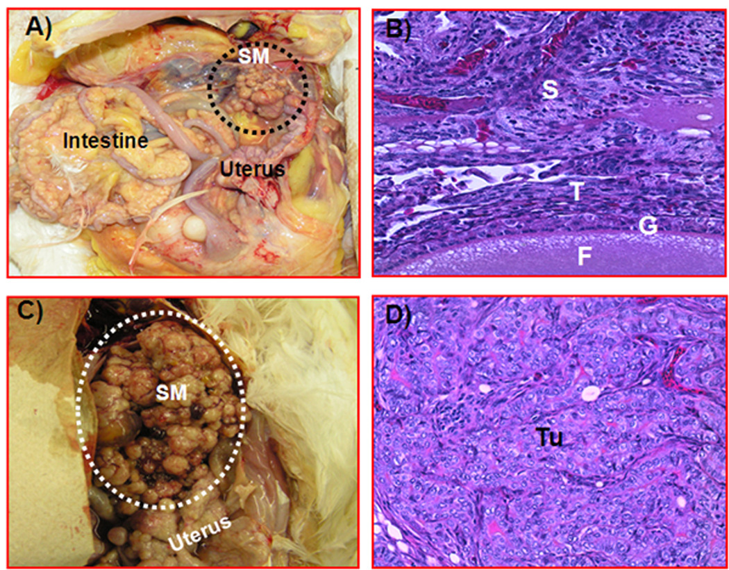
Figure 3.
Detection of ovarian tumor-associated neoangiogenesis by the postcontrast Doppler intensity of blood vessels. A flock of 150 hens were monitored for their ovarian function, and their egg-laying rates were recorded on a daily basis. Hens with low-egg laying rates (n = 46) were selected for sonography. On the basis of gray scale sonography and gross and histopathologic examinations, hens were grouped as having normal ovaries (n = 24 hens), microscopic ovarian cancer (OVCA; n = 7), early-stage ovarian cancer (n = 4), and late-stage ovarian cancer (n = 11). Postcontrast Doppler intensities of ovaries and ovarian tumors were measured by power Doppler sonography after Optison injection. Cutoff lines (mean peak Doppler intensity values of normal hens with 2 or 3 SDs) indicate the detectability of ovarian cancer by postcontrast peak Doppler intensities.

