Figure 1.
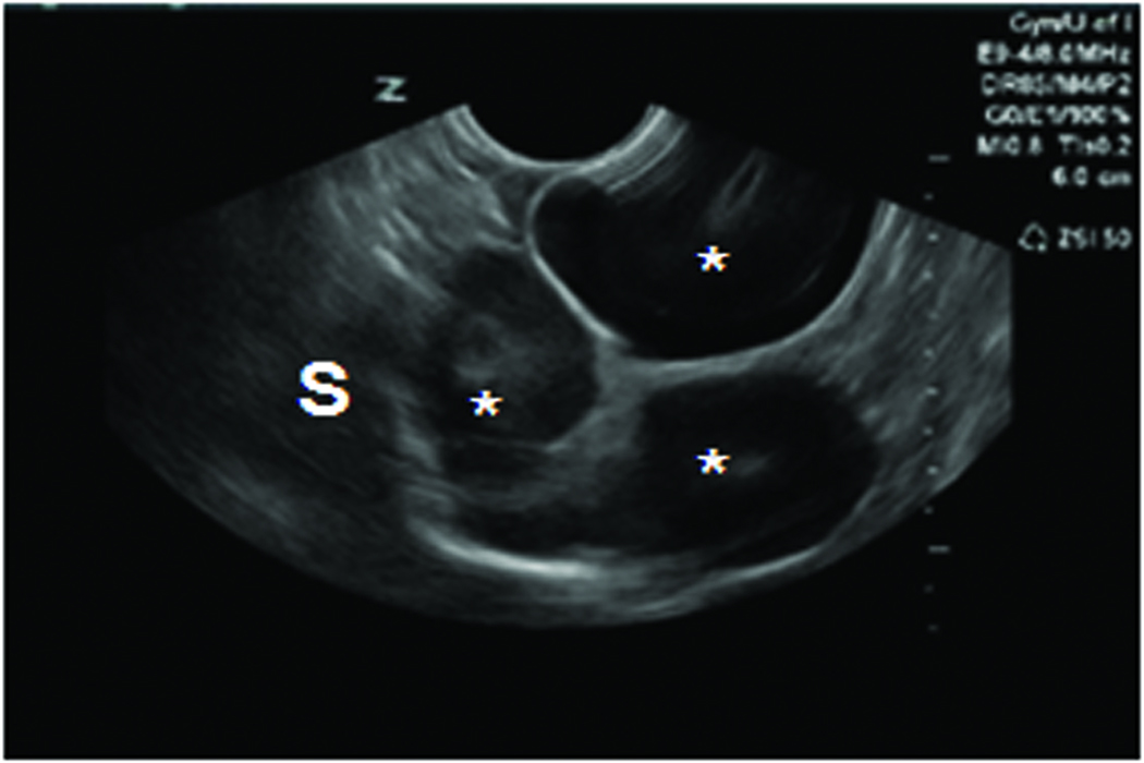
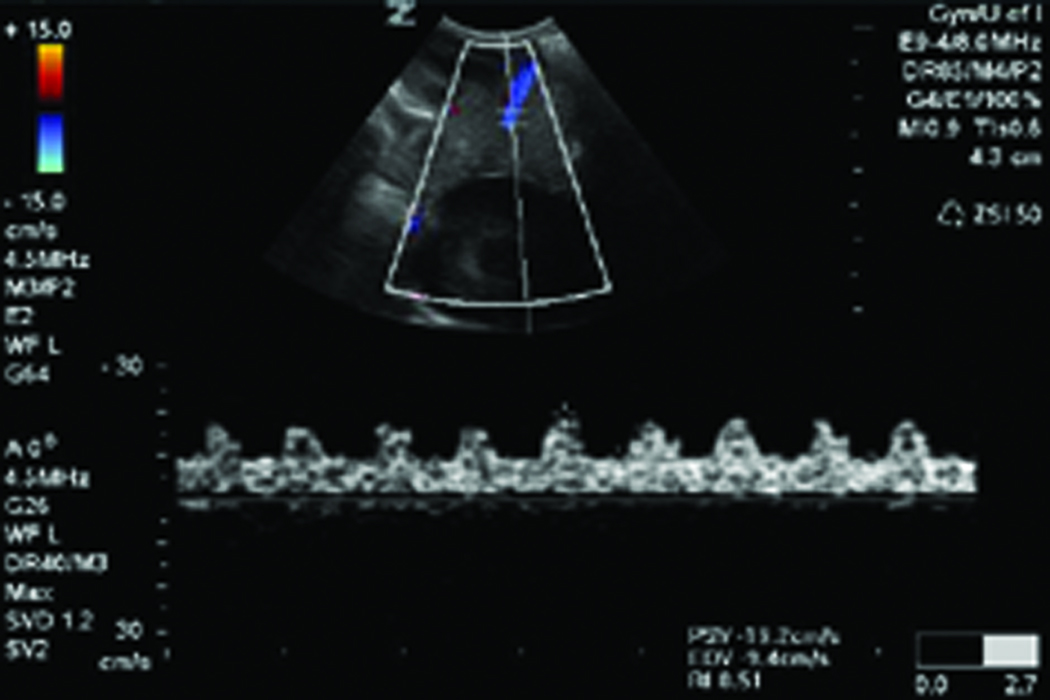
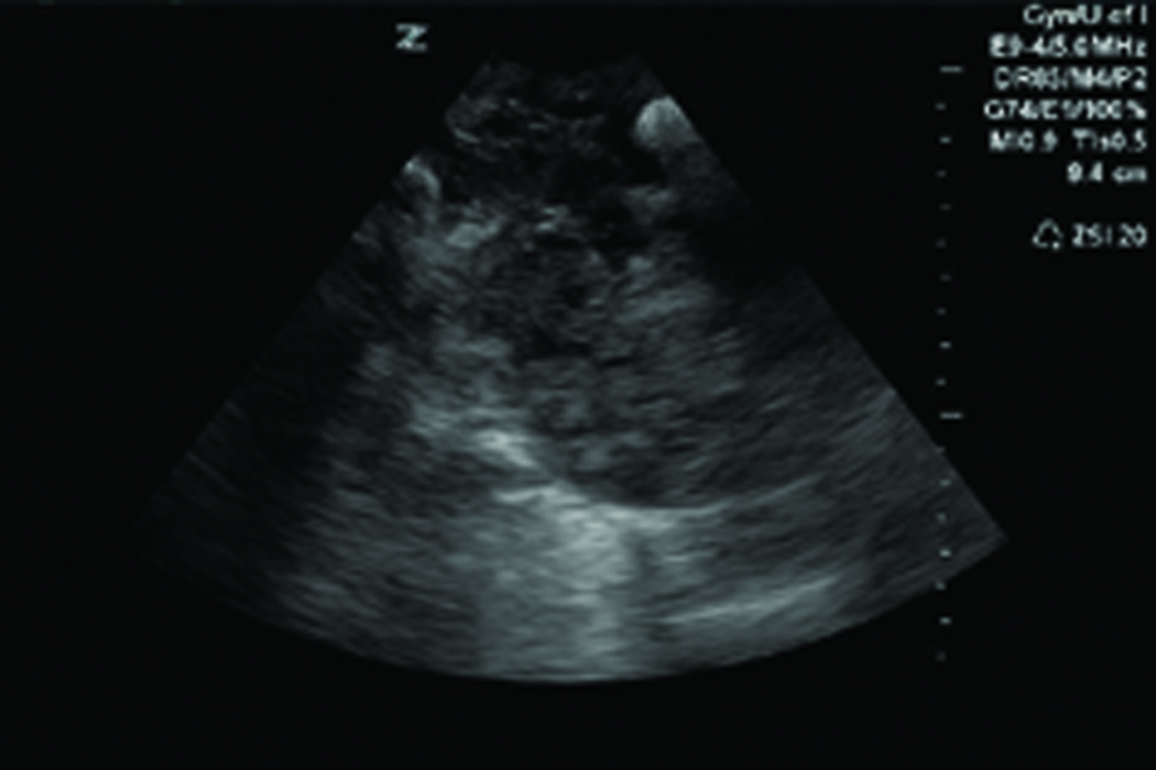
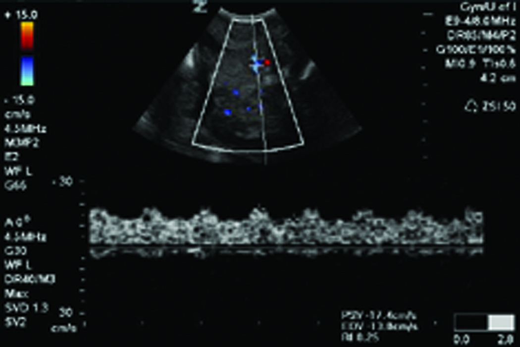
Changes in ovarian morphologic characteristics with blood flow patterns leading to tumor development in laying hens. A, Gray scale sonogram of a normal hen ovary at second scan (15 weeks after first scan). The presence of multiple preovulatory follicles (asteriks) of various sizes with no solid mass indicates the functionally normal ovary. B, Doppler sonogram of the same ovary imaged in A showing blood flows on the follicular wall of the larger preovulatory as well as small stromal follicles, suggesting active follicular growth in the ovary. C, Gray scale sonogram of the ovary of same hen shown in A at third scan (after 30 weeks from first scan). No developing follicle is seen and the ovary appears to have solid tissue masses indicating abnormal morphologic characteristics suggestive of ovarian tumor. D, Doppler sonogram of the same ovary shown in C. A central pattern of blood flow is seen on the solid ovarian mass, suggesting the presence of ovarian tumor. EDV indicates end-diastolic velocity; PSV, peak systolic velocity; and S, stroma.


