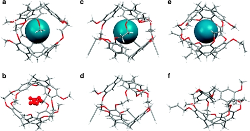
Figure 2. X-ray crystal structures of cryptophanes and inclusion complexes in side view.
(a) Cryptophane-A with Xe, and b, with water, oxygen shaded red; both in CC conformation. (c) Tripropargyl cryptophane (1), with Xe, and d, partially occupied; both in CC conformation. (e) Triallyl cryptophane (2), with Xe, in CC conformation. Xe atom is shaded blue; and f, collapsed, in CS conformation.

