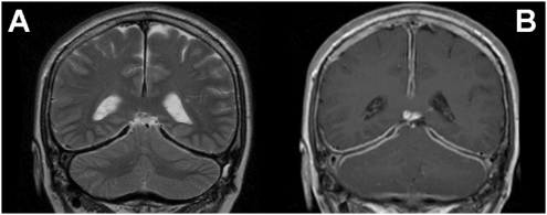
Figure 2.
Pachymeningitis (case 2). Both images clearly show the linear dural thickening and enhancement found in case 2. The differential diagnosis includes carcinomatous meningitis, intracranial hypotension, sarcoidosis, histiocytosis, idiopathic hypertrophic cranial pachymeningitis and dural sinus thrombosis. (A) Coronal T2-weighted sequence. (B) Coronal T1-weighted sequence with contrast enhancement.

