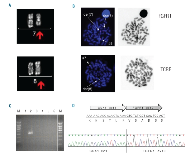
Figure 1.
Cytogenetic and molecular identification of CUX1 as a fusion partner of FGFR1. (A) Cytogenetic analysis of patient’s peripheral blood cells. The derivative chromosomes 7 and 8 are shown with the arrows. (B) Fluorescence in situ hybridization using RP11-350N15 (spectrum orange) spanning the FGFR1 locus (8p11), and RP11-556I13 (spectrum orange) and RP11-1220K2 (sybr green) flanking TCRB (7q34). Hybridization of the FGFR1 probe shows one normal signal on chromosome 8, one signal on der(7) and one signal on der(8). Hybridization of the TCRB probe confirms translocation of the distal part of 7q to 8p. (C) RT-PCR. Lanes 1 and 4: negative control (no cDNA), lanes 2 and 5: cDNA from the reported patient; lanes 3,6: cDNA from a case with CEP110-FGFR1. Forward primer in CUX1 and reverse primer in FGFR1 (Lane 1-3); forward primer in FGFR1 and reverse primer in CUX1 (lane 4–6). Lane M represents 1kb size standard. (D) Schematic representation of the identified fusion transcript. The DNA and protein sequence at the fusion border are presented. An electropherogram with the CUX1-FGFR1 fusion sequence of the PCR product is also shown.

