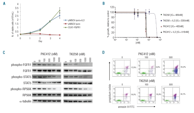
Figure 2.
In vitro assays on CUX1-FGFR1-expressing Ba/F3 cells. (A) IL-3 deprivation of Ba/F3 cells transduced with CUX1-FGFR1 resulted in transformation to growth factor independent growth. The mean growth ± SEM of 3 separate measurements over four consecutive days is presented. (B) The dose-response curves of CUX1-FGFR1-transduced Ba/F3 cells, treated with TKI258 and PKC412 for 48 h in the absence or presence of IL-3 (2ng/mL) are presented. Points represent the average results of 2 experiments performed in triplicate plotted with the curve-fitting GraphPad Prism 5 software; bars, SD. The calculated IC50 for each inhibitor is indicated. (C) Western blot analyses of CUX1-FGFR1-transformed Ba/F3 cells after treatment with PKC412 and TKI258. Phosphorylation of CUX1-FGFR1 and its downstream effectors STAT5 and RPS6K decreased with increasing inhibitor concentrations. Expression of total CUX1-FGFR1, STAT5 and RPS6K remained unaffected. (D) Effect of PKC412 and TKI258 on apoptosis of CUX1-FGFR1-expressing Ba/F3 cells after treatment for 48 h. The percentage of apoptotic plus necrotic CUX1-FGFR1-transduced Ba/F3 cells is indicated.

