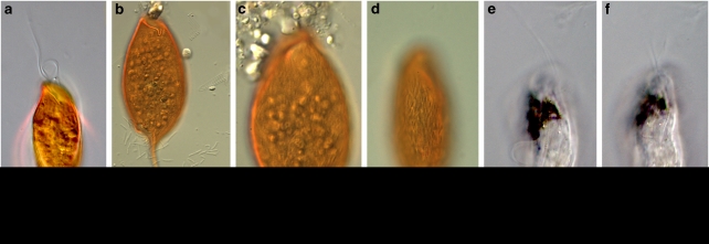
Figure 1.
Differential interference contrast (DIC) light microscope images of B. bacati and Calkinsia aureus. (a–d) C. aureus showing the epibionts that disassociate with the host almost immediately upon stress. Cells can be seen floating free of host cell in (b) and (c). (e and f) B. bacati showing black inclusions within the anterior part of the cell. Scale bar=20 μm except panel (c), which is 10 μm.

