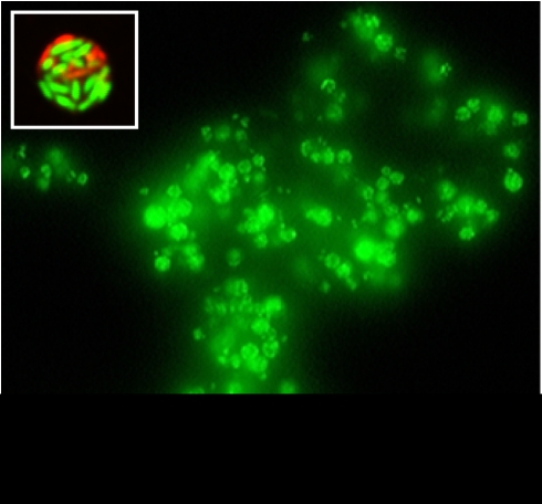
Figure 1.
Epifluorescence micrograph of a large aggregate of fecal pellets released by Tetrahymena sp. upon feeding on S. Typhimurium and stained with SYTO 9 (Invitrogen). The inset shows a single optical scan through a Live/Dead BacLight-stained fecal pellet containing live (green) and dead (red) S. Typhimurium cells. The micrograph was captured with a Leica SP5 AOTF confocal microscope (Leica Microsystems, Wetzlar, Germany).

