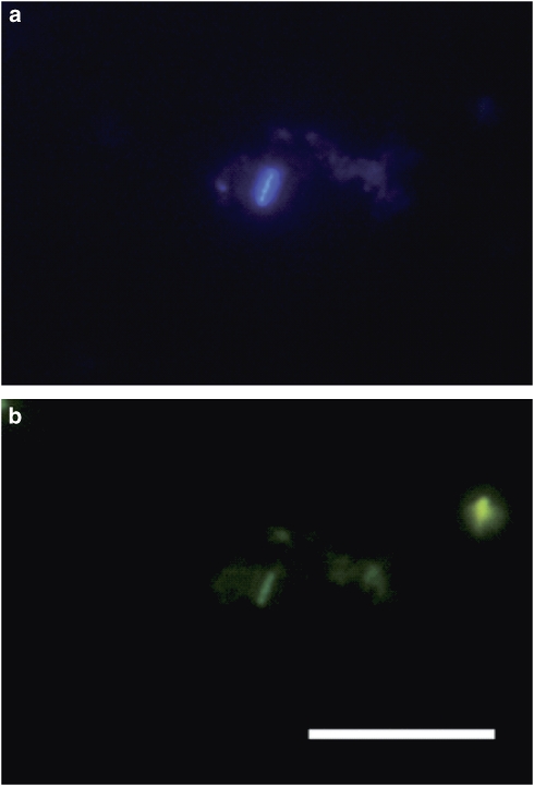
Figure 2.
Fluorescence in situ hybridization (FISH) of an ESTEC clean room sample, showing a rod shaped microorganism stained with archaeal probes. (a) Dapi stain. (b) Same section. Rod shows a positive archaeal signal, staining with an archaeal probe mixture (rhodamine green labeled). Bar: 10 μm. No signal was obtained with bacteria-specific probe mix staining in parallel (Cy3 labeled, not shown).

