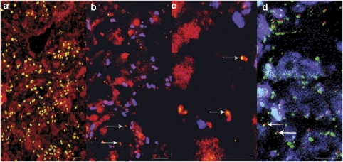
Figure 3.
Detection of sponge-associated bacteria by fluorescence in-situ hybridization (FISH). (a) Section of C. concentrica hybridized with a Cy3-labelled bacteria-specific (EUB322_CY3) probe (yellow) with the auto-fluorescent sponge tissue (red). (b) Section of C. concentrica hybridized with a Cy3-labelled δ-proteobacterium-specific probe (yellow), auto-fluorescence background (red) and larger auto-fluorescence cells (blue-purple). Arrows indicated the sponge δ-proteobacterium associated with auto-fluorescent cells (red). (c) Section of C. concentrica (enlarged) hybridized with a CY3-labelled δ-proteobacterium-specific probe (yellow). Arrows indicated the host-associated nature of the sponge δ-proteobacterium within auto-fluorescent cells (red). (d) Section of C. concentrica (enlarged) hybridized with a Cy3-labelled δ-proteobacterium-specific probe (red) and a fluorescein isothiocyanate (FITC)-labelled bacteria-specific (EUB322_FITC) probe (green). The auto-fluorescent background of sponge tissues (blue) is also shown. Arrows indicated the associated nature of the sponge proteobacterium within a putative cyanobacterial cell (green).

