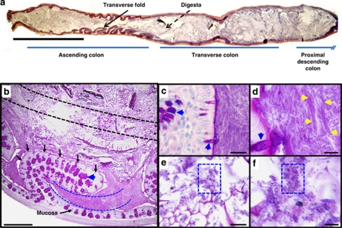
Figure 1.
A morphologically distinct population of predominately fusiform-shaped bacteria is located between the transverse folds of the mouse ascending colon. (a) Hematoxylin/eosin and (b–f) periodic acid-schiff -stained sections of the mouse colon. (a) The proximal portion of the colon (ascending colon) contains transverse folds that project into the lumen (denoted by arrow). The digesta is food particle-associated material in the central lumen (denoted as dashed arrow). (b) Methacarn-fixed section of a mouse ascending colon. The transverse fold (outlined in black arrows) emanates from the mucosa, and is lined by an epithelium that contains periodic acid-schiff-positive goblet cells (denoted as blue arrowhead). Interfold and digesta regions collected by LCM are denoted by blue and black dashed lines, respectively. (c, d) Higher-power views of interfold region. Interlacing fusiform-shaped microbes (denoted as yellow arrowheads) are abundant in this region. (e, f) Higher-power views of the digesta shows rod- and coccoid-shaped microbes (denoted as blue boxes). Bars=5 mm (a), 500 μm (b), 20 μm (c, e), 5 μm (d, f).

