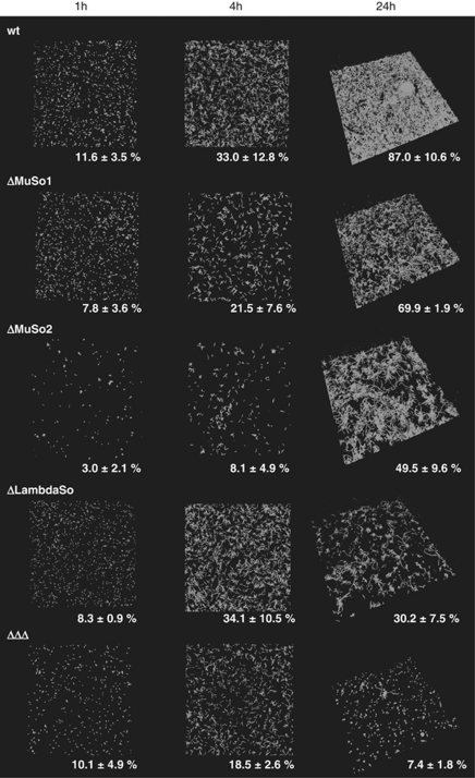
Figure 7.
Involvement of prophages MuSo1, MuSo2 and LambdaSo in biofilm formation under hydrodynamic conditions. Gfp-tagged S. oneidensis MR-1 wild-type and mutant cells were incubated in flow chambers, and biofilm formation was microscopically analyzed via CLSM after 1 (left panel), 4 (middle panel) and 24 h (right panel) of attachment. Displayed are three-dimensional shadow projections. The numbers represent the average surface coverage. The lateral edge of each micrograph is 250 μm in length.

