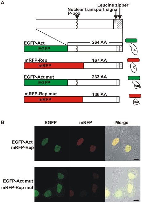
Figure 1. Expression of fluorescent-labeled CREB1 isoform proteins in HeLa cells.
A, Expression constructs. Each CREB1 activator (Act) or truncated activator (Act mut) proteins was fused with EGFP (green box), and each CREB1 repressor (Rep) or truncated repressor (Rep mut) protein was fused to tandem mRFP (red box). Within CREB1 isoform proteins, the functional domains are shown as patterned boxes. Schematic diagram of the fluorescent-labeled CREB1 isoform proteins are shown on the right side. B, Confocal microscopy images of HeLa cells transfected with expression constructs encoding EGFP- (green) or mRFP- (red) labeled CREB1 isoform proteins. CREB1 isoform proteins (EGFP-Act and mRFP-Rep, top) or truncated CREB1 proteins (EGFP-Act mut and mRFP-Rep mut, bottom) were cotransfected, and the cells were imaged 16 h after transfection. Scale bars, 10 µm.

