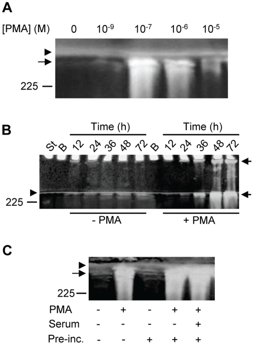
Figure 2. Synthesis of proMMP-9/CSPG in the absence and presence of PMA.
THP-1 cells were incubated in the absence or presence of PMA in serum free medium. Harvested medium was thereafter applied to Q-Sepharose chromatography and the presence of proMMP-9/CSPG was detected with gelatin zymography as described in Materials and Methods. In (A), cells were incubated for 72 h in the presence of various concentrations of PMA as indicated. In (B), cells were incubated for various time periods (as indicated) in the absence or presence of 10−7 M PMA. The samples (containing CSPG and proMMP-9/CSPG complex) from cells not exposed to PMA were five times more concentrated than the samples from the PMA treated cells when applied to the gel. In (C), cells were either incubated for 72 h in the absence (−) or presence (+) of 10−7 M PMA at serum free conditions, or pre-incubated for 3 h in the absence (−) or presence (+) of 10−7 M PMA and/or 10% fetal calf serum. After the pre-incubation, cells were washed three times in PBS and thereafter incubated in serum free medium for 72 h. Arrowhead shows the border between the separating and stacking gel, and arrows show the position of the proMMP-9/CSPG complexes. Purified proMMP-9 was used as a standard and the position of the 225 kDa homodimer form is shown at the left. The gels are representative for several similar experiments.

