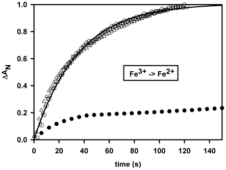
Figure 2. Anaerobic reduction.
GLB-26 (white circles), human Ngb (white triangles) and Cyt-c (white squares) by 1 mM β-NADPH under continuous light exposure (30 W deuterium lamp of the HP8453 spectrophotometer). After turning off the light or under air exposure the globin return to its initial oxidized state (no transient oxy spectrum was observed). The reduction kinetics for GLB-26 and Ngb were quite similar with a rate equal to 0.028/s, as opposed to horse heart Mb (black circles) for which two thirds of the reaction was reached after 20 mn.

