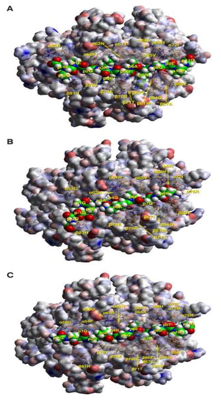
Figure 2.

Molecular simulation complexes of CII peptides with respective human MHC II alleles. A) TCR view of the chick CII peptide 286–298 (GEEGKRGARGEPG, anchors in bold, core nonamer underlined) in the groove of HLA-DQ8, after energy minimization at pH 5.4 based on the crystal structure of HLA-DQ8 (22). The α1β1 domain of the DQ302 molecule is depicted in van der Waals surface representation, with the surface atomic charges color-coded (blue, positive; grey, neutral; red, negative), and the antigenic peptide is shown in space filling form (color code: carbon, green; oxygen, red; nitrogen, blue; hydrogen, white; sulfur, yellow). Several DQ302 residues that have particular interactions with the antigenic peptide or the cognate TCR are shown in stick form with transparent van der Waals surfaces, and with the same color-code as the antigenic peptide with the exception of carbon (orange). Figure was drawn with the aid of Web Lab Viewer version 3.5 and DS Viewer Pro version 6.0, of Accelrys. TCR view of the chick CII peptides B) 53–63 (DDGEAGKPGKA, anchors in bold, core nonamer underlined) in the groove of HLA-DQ6.1, and C) 187–199 (GAQGPRGEPGTPG, anchors in bold, core nonamer underlined) in the groove of HLA-DQ6.4, after energy minimization at pH 5.4 based on the crystal structure of HLA-DQA1*0102/B1*0602 (21) Mode of depiction, colors and conventions as in 2A.
