
Fig. 1.
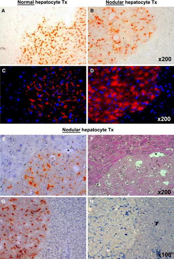
Histochemical staining for DPPIV enzyme activity (orange-rust) in clusters of normal (a) and nodular (b) hepatocytes transplanted into the liver of DPPIV-deficient host rats pre-treated with RS. Immunofluorescence labelling for DPPIV expression (red) in clusters of normal (c) and nodular (d) hepatocytes transplanted as above; DAPI (blue) identifies nuclei. Both enzyme activity and protein distribution were typically associated with the cell membrane in normal hepatocyte clusters (a, c), while they were irregular and diffuse to the cytoplasm in the expanding clusters of nodular hepatocytes (b, d). e, f Two serial cryostat sections of a nodular hepatocyte cluster stained for DPPIV expression (e, orange-rust) and with standard H&E (f). Note the presence of megalocytes in the RS-treated surrounding liver (upper-left). g, h Two serial sections of a large nodular hepatocyte cluster stained for DPPIV (g) and for the presence of CD44 marker (h); CD44-positive non-parenchymal cells are present inside the nodule, while transplanted nodular hepatocytes are negative