
Figure 3.
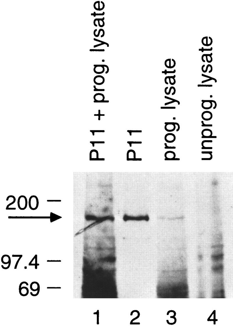
The polypeptide encoded by the cloned cDNA comigrates with endogenous hRPC155. The immunoblot was probed with the α-CSH499 antibody. (Lane 1) Mixture of 5 μl of P11 fraction (see Methods) and 5 μl of programmed reticulocyte lysate; (lane 2) 10 μl of P11 fraction; (lanes 3, 4) 5 μl of programmed and nonprogrammed reticulocyte lysate, respectively. After decay of the chemiluminescent signal, the membrane was exposed to a second film for visualization of the l[35S] methionine-labeled in vitro-translated proteins. The outline of the membrane, which was visible on both film exposures, was then used as a guide to overlay the two films. The 35S signals present in lanes 1 and 3 (but not in lanes 2 and 4) could be superimposed exactly onto the chemiluminescent signals, as expected.