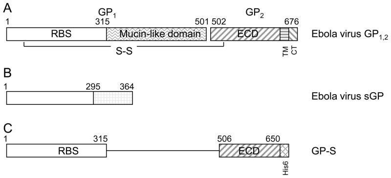
Fig. 2. Diagrammatic depiction of ebolavirus GP1,2 and antigen GP-S used for immunization.
A. full-length ebolavirus GP1,2, B. sGP; C. GP-S chimeric protein that includes aa1-315 and aa506-650 of SUDV GP1,2. The mucin-like domain, GP1/GP2 cleavage site, transmembrane domain (TM), and cytoplasmic tail (CT) were deleted, and a His6 epitope was added to the C-terminus. RBS: receptor-binding site; ECD: extracellular domain.

