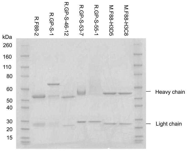
Fig. 4. Coomassie blue staining of rabbit and mouse anti-ebolavirus GP1,2 antibodies.
Each of the polyclonal antibodies was purified from sera using the corresponding antigens conjugated to beads and monoclonal antibodies were purified from the supernatant of hybridoma cells grown in serum-free medium using protein A (for rabbit monoclonals) or protein G (for mouse monoclonals) beads. Two micrograms of each antibody were resolved on SDS-PAGE gel and stained.

