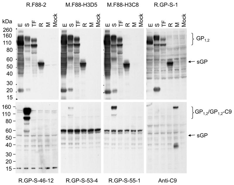
Fig. 5. Detection of various Filovirus GP1,2 by anti-ebolavirus GP1,2 antibodies using WB.
Lysate of cells that were transiently transfected to express the full-length GP1,2 of EBOV (E), SUDV (S), TAFV (TF) or the MARV (M), RESTV sGP (R), or without plasmid (Mock) were resolved on a 4-12% NuPAGE gel, and transferred to a PVDF membrane. Shown here are the bands detected after incubation with the indicated mouse or rabbit primary antibodies (indicated above or below the blots) and the corresponding horseradish peroxidase-conjugated secondary antibodies. Anti-C9 was used for detecting MARV GP1,2, which had the C9 epitope attached to its C-terminus. The size bands expected for GP1,2, sGP, and the GP-C9 fusion protein are shown on the right.

