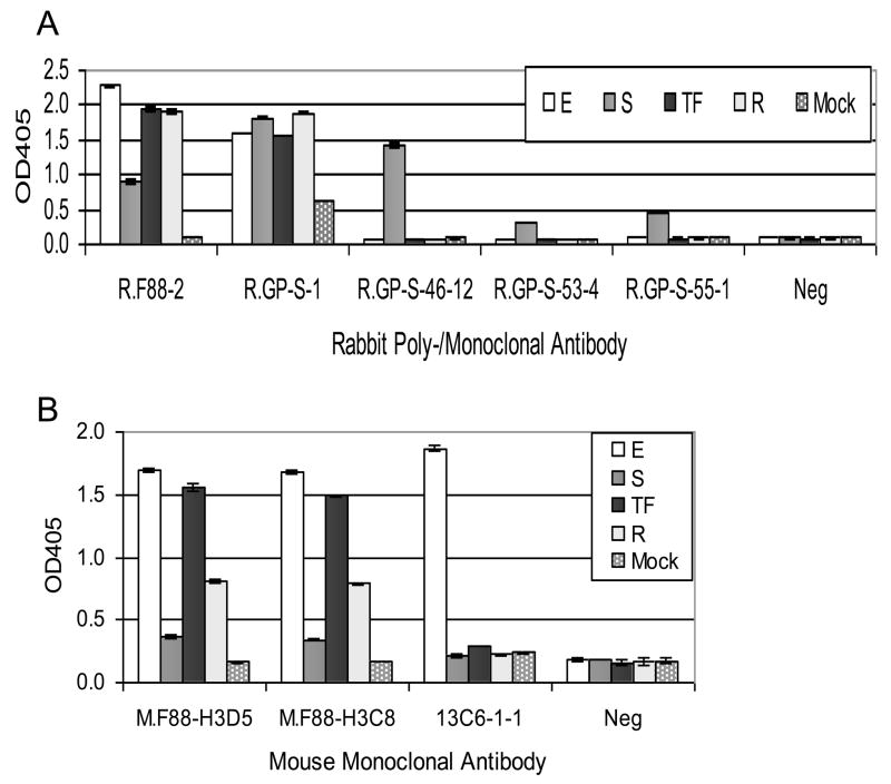
Fig. 6. Detection of various ebolavirus GP1,2 by anti-GP1,2 antibodies using sandwich ELISA.
A. To detect GP1,2 using rabbit antibodies, mouse anti-F88 monoclonal M.F88-H3D5 (2 μg/ml) was used to coat the plate and capture the indicated GP1,2 in cell lysate of transiently transfected cells. B. To detect GP1,2 by mouse antibodies, rabbit anti-F88 polyclonal R.F88-2 (2 μg/ml) was used to coat the plate and capture the indicated GP1,2. The lysates were the same as those used for WB (Fig. 5). E, EBOV GP1,2; S, SUDV GP1,2, TF, TAFV GP1,2; R, RESTV sGP. For negative controls (Neg), PBS was used in place of primary antibody, and the same horseradish peroxidase conjugated anti-rabbit IgG (A) or anti-mouse IgG (B) secondary antibody was used. Antibody 13C6-1-1 served a positive control for the assay design, by detection of EBOV GP1,2. The average OD405 ± standard deviation of triplicate samples is shown.

