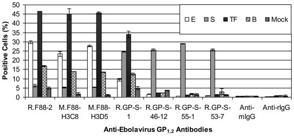
Fig. 8. Detection of various ebolavirus GP1,2 by anti-GP1,2 antibodies using flow cytometry.
COS-7 cells were transiently transfected with a plasmid that encodes GP1,2 of either EBOV (E), SUDV (S), TAFV (TF), or BDBV (B), or β-galactosidase (Mock), stained with the indicated primary antibodies (first five groups) or without the primary antibody (last two groups) and the corresponding Alexa 647-conjugated anti-rabbit or mouse IgG. Dead cells, detected positively with a stain for cell viability (YO-PRO-1), were excluded from analysis. The average percentage of positively stained cells ± standard deviation of duplicate samples is shown.

