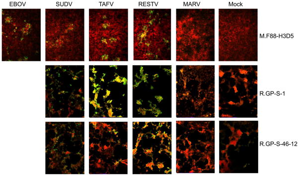
Fig. 9. Detection of filovirus-infected cells using immunofluorescence.
As described in Materials and Methods, Vero or Hela cells were exposed to different filoviruses and two days later fixed and stained with the indicated antibodies. HCS CellMask Red was for nuclear/cytoplasmic staining of all cells (shown in red). Cells that are positive for virus staining are shown in green (10X objective).

