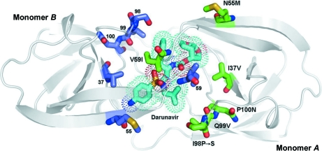
Figure 6.
The structure of 6s-98S FIV PR with DRV bound viewed along the local twofold axis of the dimer. All atoms of the mutated residues are shown (monomer A, C atoms in green; monomer B, C atoms in blue). DRV C atoms are shown in cyan, O atoms in red, N atoms in blue and S atoms in yellow. The dots represent the van der Waals surface for DRV. Monomers A and B are labeled.

