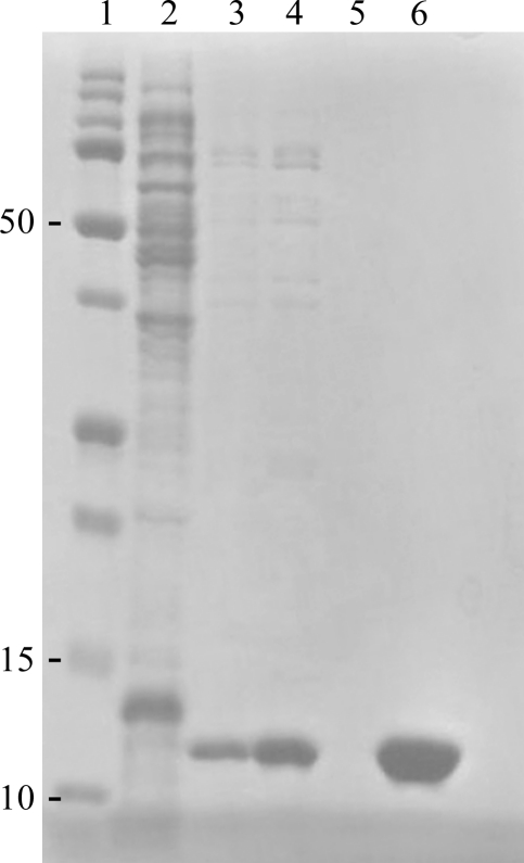
Figure 1.
Samples from recombinant FhbB purification steps as detailed in §2 were separated by SDS–PAGE (15% Tris–HCl) and the gel was stained with Coomassie Blue to visualize the proteins. Lane 1, Precision Plus molecular-weight marker (Bio-Rad); lane 2, crude cell lysate; lanes 3–5, buffer washes from the IMAC column; lane 6, pooled elution fractions.

