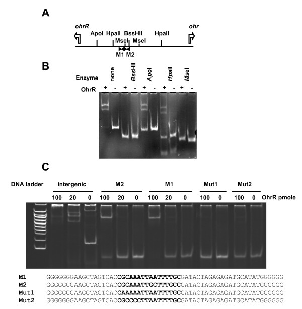
Figure 3.
Localisation of OhrR binding sites. A-Restriction map of the 113 bp ohr-ohrR intergenic region used in gel mobility shift assay. The location of the initiator codon and translation direction of ohr and ohrR is indicated by a white arrow. The position of the two palindromic binding motifs Motif 1 (M1) and Motif 2 (M2) is indicated by black arrows. B-Gel mobility shift assay of the ohr-ohrR intergenic region and of its restriction fragments produced by BssHII, ApoI, HpaII or MseI. DNA (20 pmoles) was incubated in the presence (+) or in the absence (-) of 20 pmoles of OhrR. C-Binding of OhrR to Motif 1 and Motif 2 sequences. Gel shift assay of the intergenic region and the 60 bp double strand sequences containing at their centre the genuine 17 nt corresponding to Motif 1 and Motif 2, or mutated Motif 1 with AA in place of GC (Mut1 fragment) and CCC in place of AAA (Mut2 fragment). DNA (20 pmoles) was incubated with the indicated amount of OhrR in the presence of 1 mM DTT.

