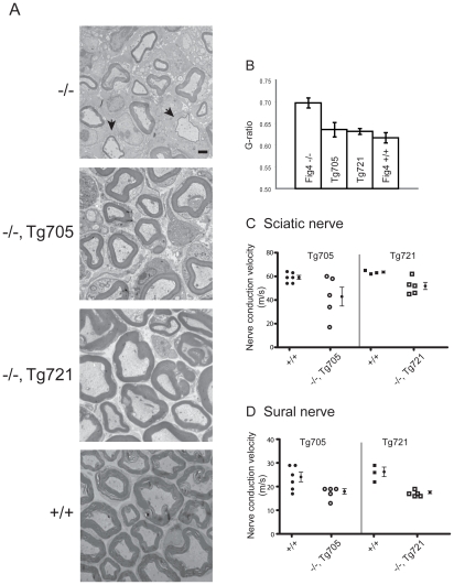
Figure 7. Rescue of peripheral nerve myelination and nerve conduction velocity in transgenic Tg705 and Tg721 lines.
A) Transmission electron microscopy of cross-sections of sciatic nerve from Fig4 mutant and wildtype mice at P21. Arrows in the top panel indicate thinly myelinated axons in Fig4−/− mice. B) Quantitation of g-ratio in sciatic nerve; higher g-ratio indicates a thinner myelin sheath. Fig4 −/− (n = 89 axons); Fig4 −/−, Tg705 (n = 181 axons); Fig4 −/−, Tg721 (n = 152 axons); Fig4 +/+ (n = 109 axons). Scale bar: 5 µm. Error bars, SEM. P<0.05 for Fig4−/− versus WT, Fig4−/− versus Tg705, and Fig4−/− versus Tg712 (Student's t-test). C, D) Nerve conduction velocity was measured in sciatic nerve and sural nerve from 4 month old unaffected Fig4−/−,Tg705 mice and 14 month old unaffected Fig4−/−,Tg721 mice (mean +/− SEM).

