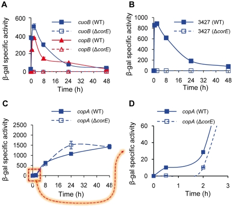
Figure 2. CorE-dependent genes.
(A) Regulation of cuoB and copB by CorE. Plasmids containing cuoB-lacZ (blue lines) and copB-lacZ (red lines) fusions were introduced into the WT (solid symbols) or the ΔcorE (open symbols) backgrounds, and incubated on CTT agar plates containing 0.6 mM CuSO4. β-gal specific activity was determined in cell extracts harvested at the indicated times. The same approach reported above was followed to study the regulation of MXAN_3427 (B) and copA (C and D) by CorE, although 0.3 mM CuSO4 was used to get an optimal difference in the copA expression levels between the WT and the ΔcorE strains at early times (panels C and D). The dashed arrow from panel C to D indicates that in panel D only the indicated part of panel C is shown. Please note the difference in the scale in each panel, and the different time course of panel D. Error bars indicate standard deviations.

