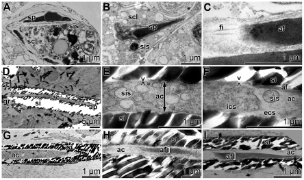
Figure 3. Longitudinal sections through spicules, showing the process of evagination of cells into the axial canals (ac) of the spicules, as elucidated by TEM.
(A and B) Intracellular onset of spicule (sp) formation in sclerocytes (scl). The growing spicules are surrounded by silicasomes (sis). (C) A developing axial filament (af), that possesses at its blunt end several filaments (fi). (D) A longitudinal section through a spicule with its axial canal (ac). The axial canal is surrounded by a silica mantel (si). The silica fragments, that occurred during cutting of the primmorphs, were partially removed. The axial canal is closed at the apex (ap) of the spicule, while it is open at the end that is associated with the sclerocyte (scl). There the growth zone of the spicule (gr) exists. (E and F) The middle part of that spicule shows in the axial canal (ac) many silicasomes (sis). Those vesicles are surrounded by a membrane, which we perceive as cell membrane (><). Furthermore, the intracellular space (ics) and the developing axial filament (af) are marked. In the extracellular space (ecs) within the axial canal (ac) a silicasome (sis) can be identified. These images were taken close to the apex of the axial canal (ac). (G) Axial canal at the apex (ac), comprising no membranous structures and no axial filament. (H and I) An axial filament (af) in the extracellular space within the axial canal (ac).

