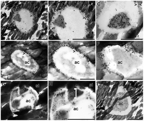
Figure 7. Immunogold labeling electron microscopy [TEM] of the axial canal of spicules.
Antibodies against silicatein (A to C), against aquaporin (D to F) and against arginine kinase (G to I) were used. (A to C) The silicatein antibodies reacted with the axial filament (af), which was surrounded by the silica mantel (si), while (D to F) the aquaporin antibodies recognized their antigens primarily at the rim of the axial canal (ac) towards the silica mantel (si). (G to I) The anti-arginine kinase antibodies reacted in a more scattered pattern with the antigen in the axial canal, primarily recognizing membranous structures. The size of all bars represents 1 µm.

