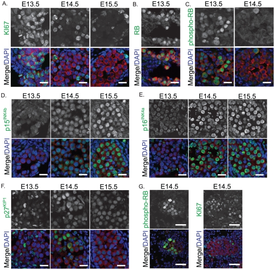
Figure 3. Immunofluorescent staining of cell cycle regulators, KI-67 and retinoblastoma (RB) in E13.5–15.5 fetal testis sections from 129T2/SvJ mice.
(A) KI-67 (green) (B) Total RB (green, E13.5 only shown) (C) phosphorylated RB (green, E13.5 and E14.5 shown), (D) p15INK4b, (E) p16INK4a and (F) p27KIP1 (G) an example of the few cords showing retained expression of phosphorylated RB and KI67 at E14.5. MVH (red) marks the germ cells and DAPI (blue) marks the cell nuclei. Scale bars; 10 um for A–F, 50 um for G.

