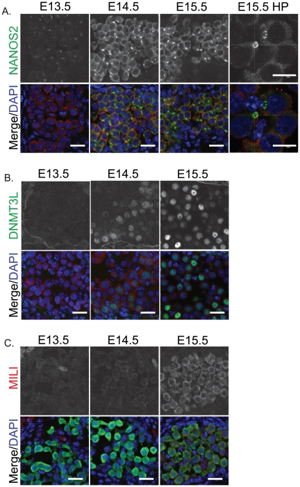
Figure 4. Male fetal germ cell differentiation markers are activated as 129T2/SvJ germ cells enter mitotic arrest.
Immunofluorescent staining of (A) NANOS2 (green), (B) DNMT3L (green), and (C) MILI (red) in E13.5–15.5 fetal testis sections from 129T2/SvJ mice. The high power image (HP) (A, far right) shows enrichment of NANOS2 protein to a cytoplasmic body that is largely MVH negative and has a NANOS2 negative core. MVH (red) marks the germ cells in A and B while GFP marks the germ cells in C. DAPI (blue) marks the cell nuclei. Scale bars; 10 um.

