
Fig. 1.
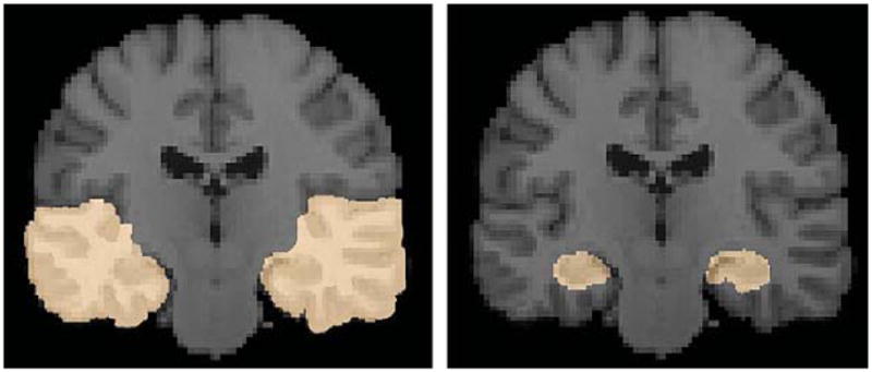
Regions of interest of the temporal lobe regions (left panel) and the hippocampus (right panel) of both hemispheres are shown. The ROIs were manually delineated on the ICBM template by a trained anatomist using the Brainsuite software program [32].