Fig. 1.
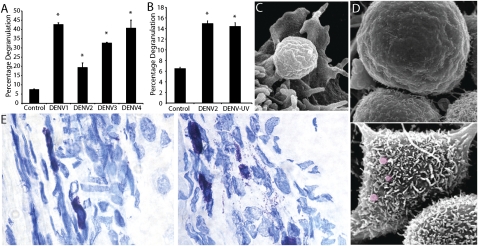
MCs degranulate in response to DENV. (A) DENV1–4 induced degranulation of RBLs. (B) Comparable RBL degranulation to live and UV-killed DENV2. Percentage degranulation in A and B was compared with spontaneous release from unstimulated MCs. *P < 0.05. (C and D) SEM of RBLs. (C) A single granule emerging from the ruffled surface of a RBL after DENV2 treatment. (D) Cell surfaces are visualized without (Upper) and with (Lower) exposure to DENV. Some extracellular granules are false-colored red. (E) Images of mouse footpad sections of control (Left) and DENV2-injected (2 × 10⁵ pfu of DENV were injected s.c.; Right) footpads. MCs in Left are fully granulated and stain metachromatically (purple), whereas MCs in Right are partially degranulated with free purple granules visible in the surrounding tissue. (Magnification: 20×.)

