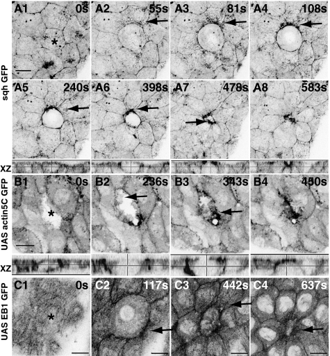
Fig. 2.
Emergent polarization of the cellular cytoskeleton in response to subcellular laser perturbation. Time-lapse confocal images of sqhGFP (A1–A8), actin5CGFP (B), and EB1GFP (C) to show actin, myosin, and microtubule dynamics upon perturbation. Arrows point to particulate streams of sqh (A1–A4), enrichment at the DC–NN interface (A5–A8), actin ruffles in the neighboring cells (B; the ablated cell is actin GFP negative), and polarized microtubule reorganization (C). Apical projections and orthogonal (XZ) views are shown. Asterisks indicate the ablated cell. See Fig. S5 for response to perturbation of tubulin GFP and microtubule organization in fixed ablated embryos. (Scale bars, 10 μm.)

