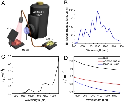
Fig. 1.
NIR II imaging. (A) Schematic of NIR II imaging setup. Anaesthetized mice are illuminated from above with 808-nm light. NIR fluorescence (1,100–1,700 nm) is filtered and imaged onto a 2D InGaAs array. (B) Fluorescence spectrum of biocompatible DSPE-mPEG functionalized SWNTs excited at 808 nm, showing several emission peaks spanning the NIR II region. (C) Absorption coefficient, μa, of water, showing the increased absorption of water in the NIR II compared to the NIR I. (D) Reduced scattering coefficient,  , for skin, adipose tissue and mucous tissue as derived in ref. 8, all showing decreased scattering with increasing wavelength.

