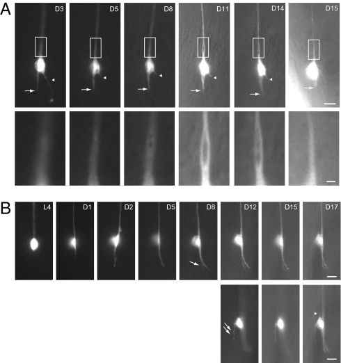
Fig. 2.
Longitudinal imaging of ALM neurons in the wild type. Epifluorescence images of individual ALM neurons at different time points over the animals’ life span (lateral view); anterior is up and ventral side is to the right. GFP is from zdIs5(Pmec-4::gfp). Ages (days of adulthood) are indicated. [Scale bar: 5 μm or 1 μm (A, Insets).] (A) The posterior process of the ALM neuron (arrow), which became slightly shorter and thicker between D3 and D5, remained static from D5 to D14, and retracted on D15. Arrowheads indicate an aberrant protrusion from the cell body, which was truncated between D3 and D5, and completely disappeared on D15. Insets highlight the development of a bubble-like structure at the proximal segment of the ALM process. The animal died on D16. (B) The posterior process of the animal grew out on D1, continued to extend between D1 and D5, and branched at D8 (single arrow). An aberrant protrusion developed from the dorsal side of the cell body at D12 (double arrows). On D17, another short protrusion developed at the anterior pole of the ALM neuron (arrowhead). (Lower) The three images were taken from different focal planes compared with their temporal counterparts in the Upper.

