Fig. 3.
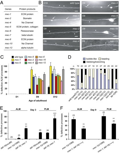
Progressive touch neuron defects in the mec mutants. (A) Summary of the mec genes and their encoded proteins. (B) Representative live GFP images (except for that of the mec-12 mutant, which is immunofluorescence image-labeled by 6–11B-1) of the ALM processes in the wild type (D16) and the mutants (D9). (Scale bar: 5 μm.) Arrows and arrowheads indicate branching and bubble-like lesions, respectively. (C) Quantification of defective ALM processes. Total numbers of ALM neurons scored (D1/D9/D12): wild type, 29/54/70; mec-1, 25/23/20; mec-2, 17/28/19; mec-4, 24/18/31; mec-5, 31/23/22; mec-6, 19/25/27; mec-9, 20/16/27; mec-10, 23/29/20; mec-12, 19/27/25. (D) Percentages of bubble-like lesions, beading, or blebbing/branching in all of the abnormal events found in the ALM neurons of wild type and the mutants. The age of the animals is specified. (E) Defects of the ALM and the PLM processes of D3 mec-1(e1066) mutants are enhanced by a daf-16(mu86) mutation. (F) Overexpression of DAF-16 from the endogenous daf-16 promoter significantly suppresses ALM and PLM defects of D9 mec-1(e1066) mutants. Error bars are SEs of proportions. **P < 0.01 or ***P < 0.001 (Fisher's exact test or two-proportion test).

