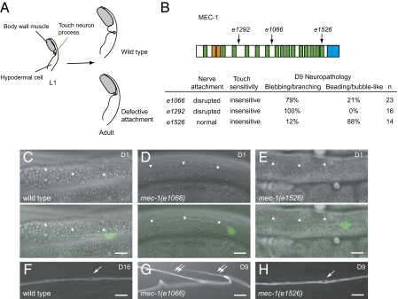
Fig. 4.
Disruption of nerve attachment or neuronal activity causes distinctive degeneration phenotypes. (Scale bar: 10 μm.) (A) Diagram of touch neuron attachment (transverse section). In wild-type L1 larvae, the process of the touch receptor neuron lies adjacent to the body-wall muscles. The nerve process later adopts the adult position where it is attached to the overlying cuticle and is thus separated from the muscles. In mutants with defective attachment, the touch neuron process remains in the juvenile position and is not attached to the epithelium. (B) MEC-1 protein structure. e1066, splice junction mutation; e1292, nonsense mutation; e1526, missense mutation. Orange, EGF domains; green, Kunitz domains; blue, the C terminus domain. Percentages of the predominant type defects (branching/blebbing or bubble-like/beading) of all axonal defects are provided. (C–E) Attachment of the ALM process in wild-type (C) and mec-1 mutants (D and E). (Upper panels) Differential interference contrast (DIC) images. (Lower panels) Overlay of DIC and GFP images. The lower borders of body-wall muscles are indicated by arrowheads. (F–H) Representative pathology of aging ALM neurons in wild-type (F) and mec-1 mutants (G and H). Single arrows: bubble-like lesions. Double arrows: branching of the nerve processes.

