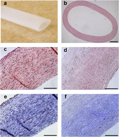
Fig. 1.
Tissue-engineered vessel from porcine SMC. (A) Porcine tissue-engineered vessel after 10 wk in culture. (B) Histology of the tissue-engineered vessel by H&E staining. H&E of the tissue-engineered vessel before (C) and after (D) decellularization with a loss of nuclei. Masson's Trichrome stain before (E) and after (F) decellularization demonstrating preservation of collagen (blue) throughout the matrix. (Scale bars: B, 500 μm; C–F, 100 μm.)

