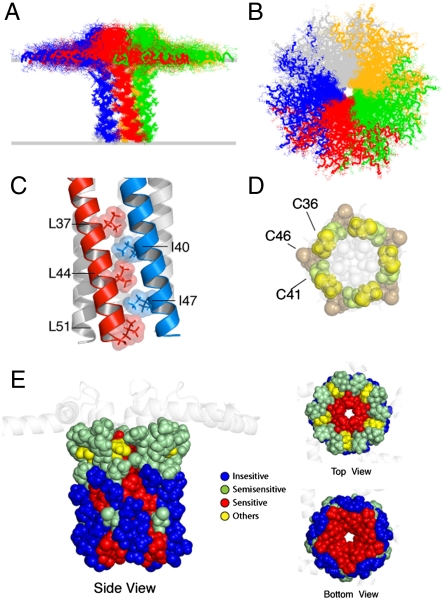
Fig. 2.
Pinwheel architecture of pentameric PLN in lipid bilayers. (A and B) PLN hybrid conformational ensemble. Overlay of the 20 lowest energy structures. The conformers were aligned using heavy atoms from residues 24 to 48. See Table S1 for structural statistics. (C) Domain II Leu/Ile zipper motif. Residues are shown using space filling model with atomic radii. (D) Spatial arrangement of the three cysteines in the PLN TM domain. The hydrophobic residues lining the inner pore are shown in light gray. Cys-41 and Cys-36 are at the interface between protomers. (E) Mapping of the mutagenesis studies on the PLN pentamer. Residues sensitive to mutation and oligomerization (1) are indicated in red. (Left) Side view of the PLN pentamer with residues from 24 to 52 shown in space filling model.

