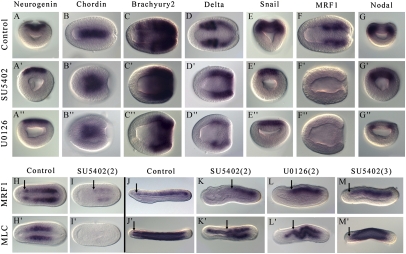
Fig. 4.
FGF and MAPK signaling inhibition induces loss of the most anterior somites. Expression patterns by whole-mount in situ hybridization of Neurogenin (A–A′′), Chordin (B–B′′), Brachyury2 (C–C′′), Delta (D–D′′), Snail (E–E′′), MRF1 (F–F′′ and H–M), Nodal (G–G′′), and MLC (H′–M′) after treatments with SU5402 (50 μM) or with U0126 (25 μM). Embryos after treatment 2 were fixed at the late gastrula stage (A–G′′), at the midneurula stage (H, H′ and I, I′), or at the premouth stage (J–L and J′–L′). Embryos after treatment 3 were fixed at the premouth stage (M, M′). A–A′′, E–E′′, and G–G′′ are blastopore views. B–B′′; C–C′′; D–D′′; F–F′′; H, H′; I, I′; and J′–M′ are dorsal views. J–M are lateral views. The most anterior limit of MRF1 and MLC is marked by a black arrow. Anterior is to the left in dorsal and lateral views, and dorsal is to the top in side and blastopore views.

