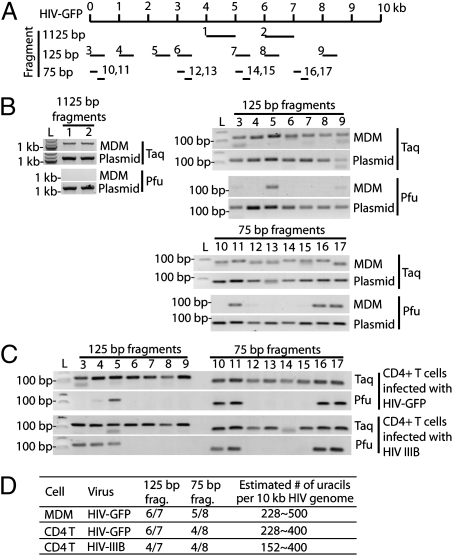
Fig. 2.
HIV DNA is heavily uracilated in infected human immune cells. (A) Schematic showing the locations of primer pairs (numbered) in the HIV-GFP genome used for uracil mapping. The numbers in this diagram correspond to labels for the gel images in B and C. (B) Taq/Pfu PCR using DNA isolated from HIV-GFP–infected MDM or plasmid DNA (as a control). (C) Taq/Pfu PCR using DNA isolated from CD4 T cells infected with HIV-GFP or HIVIIIB virus. (D) A table summarizing the findings from B and C. All primer pairs yielded expected PCR products in Taq-PCR or PCR reactions using plasmid as a template. Primer pairs that failed to yield a PCR product in Pfu-PCR were scored as uracil positive for that fragment. See text for details on how the estimated number of uracils per 10 kb HIV genome was calculated.

