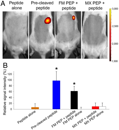
Fig. 2.
Real time measurement of PEP activity in vivo under continuous anesthesia. (A) Fluorescence imaging and (B) relative signal intensity in the stomach region 20 min after oral administration of the labeled peptide alone, the precleaved peptide, FM, or MX PEP with or without peptide. Representative rats from each set are shown in A. Color scales are identical for all pictures. In B, signals were plotted by setting the average in vivo maximum signal (precleaved peptide after 18 min) to 100%. Mean + SD, n = 6. *Precleaved peptide and FM PEP + peptide vs. all other groups (p < 0.05).

