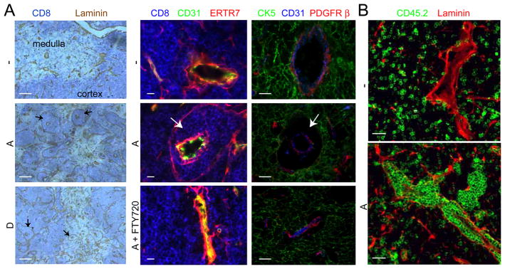
Fig. 2.
Perivascular accumulations of S1P1-transgenic thymocytes. (A) Immunohistochemical analysis of thymus sections from S1P1-transgenic and control mice with or without 24 h FTY720 treatment. Sections were stained with antibodies to detect the indicated markers. CD8 highlights the DP-rich cortical regions, ERTR7, cells associated with endothelial and epithelial basement membranes, CD31, the endothelium, CK5, medullary epithelium and PDGFRβ, pericytes. Arrows point to perivascular thymocyte accumulations. Scale bars represent 200 μm (left column) or 10 μm (middle and right columns). (B) Immunofluorescence of thymic sections from bone marrow chimeras reconstituted with a 50:50 mix of wild-type CD45.1 cells and wild-type (−) or transgenic (A) CD45.2 cells. Thymocytes of wild-type or transgenic CD45.2 origin are identified by CD45.2 staining (green). Laminin staining (red) highlights the endothelial and epithelial basement membranes. Scale bars indicate 25 μm. Data in A and B are representative of at least 3 mice analyzed in 3 experiments.

