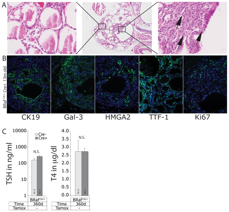
Figure 4. Thyro::CreERT2; BRafCA/+ mice develop PTC without tamoxifen administration.
A. H&E staining of histological sections of thyroid from a representative 13 month old Thyro::CreERT2; BRafCA/+ mouse that was not administered tamoxifen at low (40x, middle panel) and high power magnification (400x, left and right panels). Cells displaying abnormal nuclear cytology are indicated with arrows (right panel).
B. Immunofluorescence analysis of histological sections of thyroid from Thyro::CreERT2; BRafCA/+ without tamoxifen injection. DAPI in blue and CK19, Galectin-3, HMGA2, TTF-1, and Ki67 in green as indicated. Magnification is 200x.
C. Analysis of serum concentration of TSH and T4 from ~1 year old Thyro::CreERT2; BRafCA/+ mice that were not administered tamoxifen. N=number of mice in each group.

